 English
 Japanese
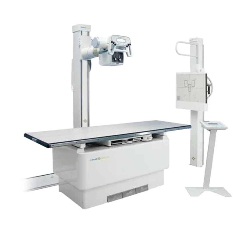
X-ray Machines
X-ray machines produce images of the inside of the body by passing controlled doses of electromagnetic radiation through tissues and capturing the attenuated beams on a detector. They are particularly effective at visualizing dense structures like bones, making them indispensable for diagnosing fractures, dislocations, and bone tumors. X-rays are also used to detect conditions like pneumonia in the lungs or foreign objects in the body. While involving a small amount of radiation exposure, X-ray imaging remains a quick, widely available, and cost-effective diagnostic tool in various medical settings.
$ 15.00
X-ray machines are fundamental diagnostic imaging tools that use electromagnetic radiation to create images of the inside of the body, most commonly to visualize bones and dense structures. When X-rays pass through the body, they are absorbed differently by various tissues; dense materials like bone absorb more radiation, appearing white on the image, while soft tissues appear gray, and air-filled spaces appear black. This technology is indispensable for diagnosing fractures, detecting lung conditions like pneumonia or tuberculosis, identifying dental problems, and evaluating joint health. Despite the use of radiation, the benefits of diagnostic X-rays generally outweigh the risks, especially with modern equipment designed to minimize exposure. X-ray imaging remains a cornerstone of medical diagnostics in emergency rooms, clinics, and hospitals worldwide due to its speed, accessibility, and effectiveness in providing critical anatomical information.